 |
Register Subscribe Login |
| 1. |  | John Dillwyn Llewelyn 1853 (ca) The Microscope Collodion negative 15.24 cm x 12.7 cm Swansea Museum, Library Swansea Museum, Library (SWASM:SM1987.845.53) The woman shown here is Thereza (1834-1926) the eldest daughter of John Dillwyn Llewelyn. Through her father's, and her own, interests in science she met and married Nevil Story-Maskelyne (1823-1911), grandson of the Astronomer Royal and professor of mineralogy at Oxford . |
| 2. |  | Thereza Llewelyn 1853-1854 (ca) Portrait of John Dillwyn Llewelyn Collodion negatve / calotype print Private collection of Noel Chanan It is not certain who took this portrait but it is unlikely to have been a self-portrait. Thereza, the eldest daughter of John Dillwyn Llewelyn, had an interest in shared science and it is possible that she took it but this is uncertain. In this photograph the setup is identical with that in the image of Llewelyn's daughter, Thereza, held by the library at Swansea Museum (SWASM:SM1987.845.53). It shows the same microscope with the same botanical specimens in front of the same books. Since the image of Thereza is larger than that of Llewelyn, we may reasonably suppose that he photographed her with his large format camera, and at the same session, she photographed him with her small format camera. Thereza notes in her diaries that everyone in the family had their own cameras. [Pers. comm. Noel Chanan, 18th April 2010] |
| 3. |  | Haack 1871 Microphotographic Camera Albumen print, mounted on cardboard Albertina © Albertina, Vienna - On permanent loan from the Austrian Federal Education and Research Institute for Graphics Carl Haack |
| 4. |  | Henry Fox Talbot 1839 Lace [Album di disegni fotogenici - The Bertoloni Album, Leaf 13 Recto] Photogenic drawing, solar microscope Metropolitan Museum of Art Harris Brisbane Dick Fund, 1936 (36.37.3) A enlargement of lace magnified 400 times with a solar microscope. |
| 5. |  | Henry Fox Talbot 1839 Lace (enlarged detail) [Album di disegni fotogenici - The Bertoloni Album, Leaf 13 Recto] Photogenic drawing, solar microscope, detail Metropolitan Museum of Art Harris Brisbane Dick Fund, 1936 (36.37.3) A enlargement of lace magnified 400 times with a solar microscope. This section of the image has been captured to show the remarkable detail of the original. |
| 6. |  | Henry Fox Talbot 1840 (ca) Photomicrograph of Moth Wings Calotype negative National Science and Media Museum |
| 7. |  | Henry Fox Talbot 1851 (ca) Photomicrograph of a crystal - study of polarized light Albumenized paper print 2 7/16 x 2 7/17 ins (6.2 x 6.2 cm) National Science and Media Museum W.H.F. Talbot Collection Probably a collaboration with William Crookes - Dr. Kelley Wilder , 8 June 2018, Observing by Polarisation, The Talbot Catalogue Raisonné - blog (Accessed: 8 June 2018) Published in "Specimens and Marvels: William Henry Fox Talbot and the Invention of Photography", Aperture, Winter 2000, Issue: 161, p.16 |
| 8. |  | Henry Fox Talbot 1851 (ca) Photomicrograph of interference patterns - study of polarized light through crystals Albumenized paper print 1 25/32 x 2 5/16 ins (4.5 x 5.9 cm) National Science and Media Museum W.H.F. Talbot Collection Probably a collaboration with William Crookes - Dr. Kelley Wilder , 8 June 2018, Observing by Polarisation, The Talbot Catalogue Raisonné - blog (Accessed: 8 June 2018) Published in "Specimens and Marvels: William Henry Fox Talbot and the Invention of Photography", Aperture, Winter 2000, Issue: 161, p.16 |
| 9. |  | Henry Fox Talbot 1840 (ca) Photomicrograph of three crystals - study of polarized light Calotype negative 2 13/32 x 2 7/32 ins (6.1 x 5.6 cm) National Science and Media Museum W.H.F. Talbot Collection, 1937-2512, Schaaf 1477 Probably a collaboration with William Crookes - Dr. Kelley Wilder , 8 June 2018, Observing by Polarisation, The Talbot Catalogue Raisonné - blog (Accessed: 8 June 2018) Published in "Specimens and Marvels: William Henry Fox Talbot and the Invention of Photography", Aperture, Winter 2000, Issue: 161, p.16 |
| 10. |  | Henry Fox Talbot 1851 (ca) Photomicrograph of interference pattern - study of polarized light through crystals Albumenized paper print 2 5/16 x 2 5/16 ins (5.9 x 5.9 cm) National Science and Media Museum W.H.F. Talbot Collection, 1937-2511/1, Schaaf 5055 Probably a collaboration with William Crookes - Dr. Kelley Wilder , 8 June 2018, Observing by Polarisation, The Talbot Catalogue Raisonné - blog (Accessed: 8 June 2018) Published in "Specimens and Marvels: William Henry Fox Talbot and the Invention of Photography", Aperture, Winter 2000, Issue: 161, p.16 |
| 11. |  | John William Draper 1844 Photomicrograph of Frog Blood Daguerreotype National Museum of American History, Smithsonian Institution Kenneth E. Behring Center, Division of Information Technology and Communications, Photographic History Collection, Image ID AF 201 |
| 12. |  | Andreas Ritter von Ettingshausen 1840, 4 March Cross section of a clematis stem Daguerreotype, whole plate Albertina Exhibited in touring exhibition: "Brought to Light: Photography and the Invisible, 1840-1900". Exhibited: Ottawa, National Gallery of Canada : "Beauty of Another Order: Photography in Science", October 1997 - January 1998. Published in: A. Thomas, Exhibition catalogue, "Beauty of Another Order: Photography in Science", New Haven & London, 1998, p.41, no.20.; A. Auer, 'Andreas Ritter von Ettingshausen (1796-1878)', History of Photography, Vol.17, No.1, Spring 1993, p.119. |
| 13. |  | John William Draper 1850 Photomicrograph of a Fly's Proboscis Daguerreotype National Museum of American History, Smithsonian Institution Behring Center, Division of Information Technology and Communications, Photographic History Collection, Image No. AFS 203 This photograph was included on the "Click! Photography Changes Everything" website (click.si.edu). |
| 14. |  | Adolphe Bertsch 1853-1857 (ca) Male Itch Mite Salt print SFMOMA - San Francisco Museum of Modern Art © San Francisco Museum of Modern Art, Members of Foto Forum |
| 15. |  | Adolphe Bertsch 1855 Antennes du Moucheron Volucelle (gross 4000 fois) Salt print 15 x 15 cm Bassenge Photography Auctions Courtesy of Bassenge, Berlin (Photography, Sale: 90, Lot: 4023, Dec 5, 2007) Adolphe Bertsch was one of the early pioneers of 19th century photography, being one of the first to make enlargements from smaller negatives. Bertsch invented the megascope which made extreme enlargements possible as early as 1860. In 1857 he published "Etudes d'histoire naturelle au microscope". |
| 16. |  | Adolphe Bertsch 1857 Bois du pin sylvestre (coupe horizontale). Gross. 1600 fs. Salt print Daniella Dangoor |
| 17. |  | John William Draper 1856 Photomicrograph of Algae Daguerreotype National Museum of American History, Smithsonian Institution Behring Center, Division of Information Technology and Communications, Photographic History Collection, Image No. AFS 146 |
| 18. |  | Alfred Donné n.d. Blood corpuscles of the frog [Donné "Atlas", fig. 8] Daguerreotype, photomicrograph Wellcome Collection Wellcome Library, London (M0011457, Museum No. 7/1950) |
| 19. |  | Léon Foucault 1844 Blood corpuscles of a frog Daguerreotype, photomicrograph 12.8 x 9.4 cm Wellcome Collection Wellcome Library, London (L0015258, Library reference no.: Iconographic Collection 578807i) |
| 20. | 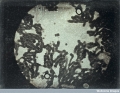 | Donné & Foucault n.d. Rouleaux of blood corpuscles Daguerreotype, photomicrograph Wellcome Collection Wellcome Library, London (L0015259, Iconographic Collections) |
| 21. |  | Léon Foucault 1844 Brewer's Yeast Daguerreotype 9.5 x 12.7 cm (3 3/4 x 5 ins) Société Française de Photographie This plate was included in the exhibition "The Dawn of Photography: French Daguerreotypes, 1839-1855". |
| 22. | 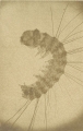 | Alois Auer 1853 (ca) [Microscopic view on an insect] Albumen silver print Metropolitan Museum of Art Rogers Fund, 1918 (18.17.1.217) MOMA includes the note: This information may change as the result of ongoing research. (5 April 2010) |
| 23. |  | Donné & Foucault 1855 Fig.40 - thin disc of cow's milk. The 120th of an inch in diameter, magnified 400 times in its linear and 160000 times in its superficial dimensions. Book page Google Books The Museum of Science and Art (London: Walton and Maberly, 1855), Volume 6, edited by Dionysius Lardner, p.97. The plate is discussed on p.106. 87. In fig. 40, p. 97, we have given the appearance presented by a thin disc, the 120th of an inch in diameter, of common cow's milk magnified 400 times in its linear, and therefore 160000 times in its superficial dimensions, engraved from a daguerreotype by MM. Donne and Foucault. |
| 24. |  | W. & F. Langenheim n.d. The Lord's Prayer - Containing 268 letters in a space the one thousand part of a square inch. and almost invisible by the naked eye. Made by W. F. Langenheim, Philadelphia Photomicrograph, slide Private collection of Herbert C. McKay Photographs prepared by Herbert C. McKay and kindly provided by his grandson Maurice M. Greeson (19 June 2012). |
| 25. |  | W. & F. Langenheim n.d. The Lord's Prayer - Containing 268 letters in a space the one thousand part of a square inch. and almost invisible by the naked eye. Made by W. F. Langenheim, Philadelphia Photomicrograph, slide, enlarged Private collection of Herbert C. McKay Photographs prepared by Herbert C. McKay and kindly provided by his grandson Maurice M. Greeson (19 June 2012). |
| 26. |  | Alfred Reeves n.d. Photograph. We Praise Thee O Lord Painted by Barraud. 8. A. Reeves, Photo. Microscope slide Private collection of Professor Brian Stevenson, Ph.D. Original photograph provided by Reg Porter. A rare microphotograph bearing A. Reeves' name spelled out. |
| 27. |  | Henry Hering 1862, 31 May The Kings and Queens of England, From the Conquest to Queen Victoria Advertisement Private collection of Professor Brian Stevenson, Ph.D. The photograph includes the 83 monarchs ruling England from the Norman Conquest to Queen Victoria. Comments by Prof. Brian Stevenson Henry Hering issued a cdv-sized version of the kings and queens montage in 1862. The illustrated advertisement is from the May 31, 1862 issue of The Bookseller, and indicates when Hering began selling these cards. Note that Reeves' slides with this image were described in 1859. Thus, it is not clear whether Hering produced this montage 3 years before he published the cdv, if Reeves produced the montage and later sold the rights to Hering, or if a third person originally made it. Noting that Hering claimed copyright for the cdv image but the Reeves microphotographs do not mention copyright, I think the last two possibilities are more likely |
| 28. |  | Henry Hering 1862 The Kings and Queens of England, From the Conquest to Queen Victoria Carte de visite Private collection of Professor Brian Stevenson, Ph.D. The photograph includes the 83 monarchs ruling England from the Norman Conquest to Queen Victoria. Comments by Prof. Brian Stevenson Henry Hering issued a cdv-sized version of the kings and queens montage in 1862. The illustrated advertisement is from the May 31, 1862 issue of The Bookseller, and indicates when Hering began selling these cards. Note that Reeves' slides with this image were described in 1859. Thus, it is not clear whether Hering produced this montage 3 years before he published the cdv, if Reeves produced the montage and later sold the rights to Hering, or if a third person originally made it. Noting that Hering claimed copyright for the cdv image but the Reeves microphotographs do not mention copyright, I think the last two possibilities are more likely |
| 29. |  | Alfred Reeves n.d. Photograph. The Kings & Queens of England, From the Conquest to Queen Victoria. Contains 85 Portraits. A.R. Microscope slide Private collection of Professor Brian Stevenson, Ph.D. Original photographs courtesy of David Evans. The original photograph which was copied for this photomicrograph was by Henry Hering (1814-1893). See the carte de visite version from 1862 in the collection of the National Portrait Gallery, London (NPG Ax131392). Thanks to T. Max Hochstetler for passing on this information (email to Alan Griffiths, 16 June 2011). Contemporary comments: "Mr. Alfred Reeves has recently forwarded to us a specimen of one of those minute pictures, which consists of a plate containing the portraits of kings and queens of England since the time of the Conquest. Here, on a space not larger than 1/16 of an inch square, may be perceived a miniature "National Portrait Gallery" with a portrait of every king and queen surrounding her Majesty, who is properly made the centre figure of the interesting group." ("Micro-Photography" The Photographic News, Volume 2, March 18, 1859, p.15) |
| 30. |  | John Charles Stovin 1858-1862 Examples of microphotograph slides by J.C. Stovin. Microphotograph slides Private collection of Professor Brian Stevenson, Ph.D. The majority of his slides bear one, pale brown label and the initials "JS". As Warren (2006) pointed out, the use of two initials on the "JS" labels achieves artistic balance. Others carry two yellowish labels and the initials "JCS". Another producer of microphotographs, the as-yet unidentified "EM" used similar labels, and may have been a colleague of Stovin's, they may have copied each other, or simply patronized the same printer. |
| 31. |  | John Charles Stovin 1862 (exhibited) Two microphotographs with titles that match descriptions of full-sized photographs displayed by Stovin at the 1862 London Exposition Microphotograph slides Private collection of Professor Brian Stevenson, Ph.D. The other nine exhibited photographs were of Government Offices, All Souls Church, Statue of the Duke of Wellington, Trafalgar Square, Somerset House (two different views), The Tower, Westminster Hospital, and Houses of Parliament. According to Nicol (1881), Stovin expanded this series of microphotographs to 36 different views. |
| 32. |  | John Charles Stovin 1858-1862 Examples of Stovin's microphotographs Microphotograph slides Private collection of Professor Brian Stevenson, Ph.D. Clockwise from top left: "Her Majesty the Queen" (Victoria), "The Kings and Queens of England, from Egbert to Victoria", "His Royal Highness Prince Albert" (this exists in two label variants, with the word 'late' added after Prince Albert's death in 1861), and "H.R.H. The Prince of Wales" (who became King Edward VII upon Victoria's death in 1901). The kings and queens montage may have been a response to an acclaimed microphotograph produced by competitor Alfred Reeves, which showed pictures of English sovereigns from the Norman Conquest (1066) until Victoria. Stovin outdid Reeves by going further back in time to Egbert, who ruled 802-839. |
| 33. |  | John Charles Stovin 1858-1862 Her Majesty the Queen (Victoria) Microphotograph slide Private collection of Professor Brian Stevenson, Ph.D. |
| 34. |  | John Charles Stovin 1858-1862 His Royal Highness Prince Albert Microphotograph slides Private collection of Professor Brian Stevenson, Ph.D. This exists in two label variants, with the word 'late' added after Prince Albert's death in 1861), |
| 35. |  | John Benjamin Dancer n.d. The Micrograph Viewer for micrographs Interencheres - La Gallerie de Chartes Collection Henry Koilski (Galerie de Chartres, Auction, 9 October 2011, Lot: 727) Petite lorgnette en ivoire avec petite vues. L'ensemble dans un écrin. |
| 36. |  | Oliver Wendell Holmes n.d. Photomicrograph of "The Declaration of Independence" Google Books Oliver Wendell Holmes, "VII. Mechanism in Thought and Morals" IN Pages from an Old Volume of Life - A Collection of Essays 1857-1881, (Boston: Hoghton, Mifflin and Company, 1883), p.298. I have a glass slide on which is a minute photographic picture, which is exactly covered when the head of a small pin is laid upon it. In that little speck are clearly to be seen, by a proper magnifying power, the following objects: the Declaration of Independence, with easily-recognized facsimile autographs of all the signers; the arms of all the original thirteen States ; the Capitol at Washington; and very good portraits of all the Presidents of the United States from Washington to Polk. These objects are all distinguishable as a group with a power of fifty diameters: with a power of three hundred, any one of them becomes a sizable picture. You may see, if you will, the majesty of Washington on his noble features, or the will of Jackson in those hard lines of the long face, crowned with that bristling head of hair in a perpetual state of electrical divergence and centrifugal self-assertion. Remember that each of these faces is the record of a life. |
| 37. |  | Adolphe Neyt 1865 (ca) Photomicrograph of a flea Albumen print 19.8 x 15.0 cm George Eastman Museum Record Id: 1977:0638:0015 |
| 38. |  | Arthur E. Durham 1863 (ca) Three Mites Albumen print National Science and Media Museum The Royal Photographic Society, Ref Number: 2003-5001/2/23760/1 |
| 39. |  | Arthur E. Durham 1865 (ca) Photomicrograph of a Fly Albertina |
| 40. |  | Arthur E. Durham 1863-1864 Photomicrograph of a Flea Albumen print National Science and Media Museum © Royal Photographic Society Collection, National Media Museum, Bradford, UK, purchased with the assistance of the Art Fund |
| 41. |  | Albert Moitessier 1866 Photo-micrographs Book plate Google Books A. Moitessier La Photographie, Appliquée aux Recherches Micrographiques (Paris, J. B. Bailliereet Fils, 1866) |
| 42. |  | Jules Girard 1869 Diatomées groupées Heliogravure Google Books Jules Girard "La Chambre Noire et le Microscope" (Paris: F. Savy, 1869), p.55, fig.5 |
| 43. |  | J. Dunlop (English) 1870 Eye of Indian Gad fly. Magnified 100 diameters Albumen print Private collection of Brad Feuerhelm |
| 44. |  | Joseph Janvier Woodward 1871 (ca) Small Artery and Capillaries from Lung of Frog [Report to the Surgeon General of the United States Army on an improved Method of Photographing Histological Preparations by Sunlight" - Issued by the U.S. War Department Surgeon General's Office - Plate No. 2 (of 9).] Albumen print, photo-micrograph 6 x 6 in (16x16 cm) Christopher Wahren Fine Photographs Courtesy of Christopher Wahren Fine Photographs (hb72.365) On fancy mount 14 x 11 inches (35x28 cm) with gilt imprint "WAR DEPARTMENT, Surgeon General's Office, Army Medical Museum… by J. J. WOODWARD, Asst. Surg., U.S.A. / By order of the Surgeon General." With pasted-on printed label reading "SMALL ARTERY AND CAPILLARIES FROM LUNG OF FROG. The preparation was injected with a dilute silver solution and subsequently stained with carmine. Magnified 500 diameters. Negative No. 365, New Series." |
| 45. |  | Unidentified photographer / artist 1880 Saccharomycetes and Schizomycetes (Nageli) Book illustration Google Books Dr. Antoine Magnin The Bacteria (Boston: Little, Brown, and Company, 1880) Saccharomycetes and Schizomycetes (Nageli), developed in urine (of yellow-fever patient) exposed in laboratory of the Yellow-fever Commission, Havana, July, 1879. Reproduced by permission of the National Board of Health. Fig. 1. Photo-micrograph made with Beck's 1/5 in. objective and Tolles's amplifier. 400 diameters. |
| 46. |  | George E. Davis 1881 Proboscis of Fly Woodburytype, magazine illustration Google Books The Northern Microscopist, No.4, April 1881, plate V. Accompanies the article on "Photo-Micrography" by George E. Davis. |
| 47. |  | Frederick H. Evans n.d. Microscopes slide of "Sponge Spicules" (left) and "Pulex Iritans (sic) - Flea" (right) with signatures Microscope slides Private collection of Professor Brian Stevenson, Ph.D. Two microscope slides bearing labels with Frederick Evans' signature. The left slide, of a flea, also carries a descriptive label in the same hand, and was presumably made by Evans. The right slide, a papered arrangement of sponge spicules, bears a specimen label with a very different style of handwriting. It is not known whether Evans bought the slide through G. Rogers, or if Rogers disposed of it after Evans had owned the slide. Enlargements of Evans' signature are included to assist readers in comparing the slides' signatures with those on photographs by Frederick Evans. http://microscopist.net/ (Accessed: May 14, 2010) Brian Bracegirdle's Microscopical Mounts and Mounters (Oxford, Seacourt Press, 1998) illustrates a slide with an Evans label on plate 15D, with a cover paper indicating that it was made by J. & T. J. (Jones). Bracegirdle interpreted the scrawled handwriting to be "F N Evans." |
| 48. | 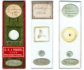 | Frederick H. Evans n.d. Microscopes slide of "Sponge Spicules" (left), "Tooth of Shark" (center) and "Pulex Iritans (sic) - Flea" (right) with signatures Microscope slides Private collection of Professor Brian Stevenson, Ph.D. Three microscope slides bearing labels with Frederick Evans' signature. The left slide, of a flea, also carries a descriptive label in the same hand, and was presumably made by Evans. The right slide, a papered arrangement of sponge spicules, bears a specimen label with the handwriting of professional slide-maker John Barnett. It is not known whether Evans bought the slide through G. Rogers, or if Rogers disposed of it after Evans had owned the slide. The maker of the center slide is not known. |
| 49. |  | Frederick H. Evans 1914 (album) Spine of Echinus (title incomplete) Photomicrograph, on album page George Eastman Museum 1973:0250:0042a. From the album "Photo-micrographs by Frederick H. Evans. Negatives and Silver prints made before 1886, other prints in Satista platinotype in 1914" held at George Eastman House. |
| 50. |  | Frederick H. Evans 1914 (album) Spine of Echinus (title incomplete) Photomicrograph George Eastman Museum 1973:0250:0042a. From the album "Photo-micrographs by Frederick H. Evans. Negatives and Silver prints made before 1886, other prints in Satista platinotype in 1914" held at George Eastman House. |
| 51. |  | Frederick H. Evans 1914 (album) Photomicrograph Photomicrograph, on album page George Eastman Museum GEH#1973:0250:0042b From the album "Photo-micrographs by Frederick H. Evans. Negatives and Silver prints made before 1886, other prints in Satista platinotype in 1914" held at George Eastman House. |
| 52. |  | Frederick H. Evans 1914 (album) Photomicrograph Photomicrograph George Eastman Museum GEH#1973:0250:0042b From the album "Photo-micrographs by Frederick H. Evans. Negatives and Silver prints made before 1886, other prints in Satista platinotype in 1914" held at George Eastman House. |
| 53. |  | Frederick H. Evans 1887 (ca) Fr: Sec: Spine of Echinus x. 40 Platinum print 4 3/4 x 4 11/16 ins (12.1 x 11.9 cm) (image) 13 5/8 x 10 5/16 ins (34.6 x 26.2 cm) (mount) Philadelphia Museum of Art Purchased with funds contributed by Dorothy Norman, 1973, 1973-197-65 Philadelphis Museum of Art accompanying text [Accessed: 24 Oct 2010] Unlike many beginning photographers of the nineteenth century who experimented with straightforward portrait or landscape compositions, Evans's earliest trials with photography involved minute organic matter and required the use of a microscope. His complicated "photo-microgram" process allowed him to capture the intricate structures of objects including a water beetle's eye, tiny sea shells, and this section of a sea urchin's spine. Although classified as scientific rather than artistic imagery by the Photographic Society of Great Britain, this photo-microgram demonstrates Evans's ability to delineate the magnificence of organic patterns and presage his photographs that depict the structural beauty of cathedrals. |
| 54. |  | Unidentified photographer / artist 1887 Bacillus anthracis Book illustration Google Books Edgar M. Crookshank Photography of Bacteria (New York: J.H. Vail & co., 1887), Plate VII, Photo II. Enlargement from a negative taken with a 1/25 hom, imm. Of Powell and Lealand. From a section of kidney of a mouse which had died after inoculation with Bacillus anthracis. Showing a glomerulus injected with the bacilli. Stained by the method of Gram. X 1500. |
| 55. |  | Unidentified photographer / artist 1887 Proteus Zenkeri Book illustration Google Books Edgar M. Crookshank Photography of Bacteria (New York: J.H. Vail & co., 1887), Plate IV, Photo I. Enlargement from a negative taken with Zeiss' DD. From a cover-glass impression-preparation of Proteus Zenkeri. The preparation was stained a very faint violet with methyl violet, and is represented in brown to illustrate the employment of a brown pigment in the autotype process. X 500. |
| 56. |  | Unknown (Great Britain) 1890 (ca) Microphotograph of a tick Cyanotype 2.75 x 3 ins Archive Farms |
| 57. |  | Wilson A. Bentley 1895 (ca) Snowcrystal Albumen print 3 x 4 in (7.5 x 10 cm) Swann Galleries - New York Courtesy of Swann Galleries (Auction, Dec 7, 2006, #2097, Lot 372) |
| 58. | 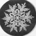 | Wilson A. Bentley 1890 Snowflake Study Albumen print 3 x 3 ins Smithsonian Institution Archives Image No. RU 31 Box 12 Folder 17 This image was used as an illustration in the Kenneth G. Libbrecht "Photography changes natural phenomena into iconic images" story on the Smithsonian - "Click! Photography changes everything" website (click.si.edu). |
| 59. |  | Richard Neuhauss 1892-1893 Snow crystals and ice structures Gelatin silver print 16.7 x 10,8 cm Feroz Gallery This photograph was included in the exhibition "Dr. Richard Neuhauss - Winter 1892" (Feroz Gallery, Bonn, Germany, 2011). |
| 60. |  | Richard Neuhauss 1892-1893 Snow crystals and ice structures Gelatin silver print 16.2 x 10.6 cm Feroz Gallery This photograph was included in the exhibition "Dr. Richard Neuhauss - Winter 1892" (Feroz Gallery, Bonn, Germany, 2011). |
| 61. |  | Richard Neuhauss 1892-1893 Snow crystals and ice structures Gelatin silver print 10.5 c 16.2 cm Feroz Gallery This photograph was included in the exhibition "Dr. Richard Neuhauss - Winter 1892" (Feroz Gallery, Bonn, Germany, 2011). |
| 62. |  | Unidentified photographer / artist 1905 (publication) 1896 (copyright) Fig.67 Ink-Crystals, as seen through a microscope Book illustration This work is out of copyright Published in Photographic Amusements including a Description of a Number of Novel Effects Obtainable with the Camera by Walter E. Woodbury (New York: The Photographic Times Publishing Association, 1905) From the Literay Gazette. |
| 63. |  | Arthur Wells Bawtree 1900 (ca) Wing of a Gnat, Magnified 40 Times Gelatin silver print 3 in (7.62 cm) x 6 in (15.24 cm) Robert Tat Gallery Courtesy of Robert Tat Fine Photographs (www.roberttat.com - #1339) |
| 64. |  | George Hook Rodman (1861-1933) 1900 (ca) Hairs on wing of a fly x 450 Gelatin silver print National Science and Media Museum The Royal Photographic Society, Ref Number: 2003-5001/2/20192 |
| 65. |  | Laure Albin-Guillot 1929 (ca) Micrographie Tirage argentique 22 x 28 cm Photo Verdeau |
| 66. |  | Carl Strüwe 1933 Stinging Nettle - Urtica - Stinging hairs (Urbild der Abwehr) Gelatin silver print, Microphotograph 35:1 24 x 18.5 cm Carl-Strüwe-Archiv © Carl-Struwe-Archiv, Bielefeld, Germany (STR-1-097) |
| 67. |  | Sondra Barrett 1979 Frangelico, Sweet Italian, Hazelnut Liqueur [Microscopic wine crystals] 13 x 19 3/8 Barry Singer Gallery |