| 1. |  | Unknown creator 2007 (uploaded) DNA molecule Rendered image, software generated Creative Commons - Wikipedia |
| 2. |  | Unknown creator 2006, 21 November DNA repair Rendered image, software generated Creative Commons - Wikipedia DNA damage, due to environmental factors and normal metabolic processes inside the cell, occurs at a rate of 1,000 to 1,000,000 molecular lesions per cell per day. A special enzyme, DNA ligase (shown here in color), encircles the double helix to repair a broken strand of DNA. DNA ligase is responsible for repairing the millions of DNA breaks generated during the normal course of a cell's life. Without molecules that can mend such breaks, cells can malfunction, die, or become cancerous. DNA ligases catalyse the crucial step of joining breaks in duplex DNA during DNA repair, replication and recombination, and require either Adenosine triphosphate (ATP) or Nicotinamide adenine dinucleotide (NAD+) as a cofactor. Shown here is DNA ligase I repairing chromosomal damage. The three visable protein structures are:
It is likely that all mammalian DNA ligases (Ligases I, III, and IV) have a similar ring-shaped architecture and are able to recognize DNA in a similar manner. (See:Nature Article 2004, PDF)
|
| 3. |  | Kieran Maher 2006, 30 October 3D VR of a CT scan: orthogonal cropping frame OsiriX Creative Commons - Wikipedia |
| 4. |  | Milorad Dimic MD (Intermedichbo, Nis, Serbia) 2009, 16 August Morbus Buerger CT angiogram Creative Commons - Wikipedia |
| 5. |  | Unknown creator 2009, 27 October This is a computer-enhanced fMRI scan of a person who has been asked to look at faces. The image shows increased blood flow in the part of the visual cortex that recognizes faces. Computer-enhanced fMRI scan Creative Commons - Wikipedia http://www.nlm.nih.gov/hmd/emotions/frontiers.html Public domain US government |
| 6. |  | Fuchs, Daniel & Geo n.d. Fetus, 8 months, with tumor [Conserving Humans] Cibachrome print Stephen Bulger Gallery |
| 7. |  | Ed Uthman 2000 (ca) Human Embryo (7th week of pregnancy) Colour film Creative Commons - Wikipedia |
| 8. |  | Unidentified photographer / artist n.d. A scoliotic spine National Museum of Health and Medicine National Museum of Health and Medicine |
| 9. |  | Unidentified photographer / artist n.d. Egg surrounded by sperm National Museum of Health and Medicine National Museum of Health and Medicine |
| 10. |  | Unidentified photographer / artist n.d. The new M2A Capsule, a pill endoscope, created by Given Imaging Ltd National Museum of Health and Medicine National Museum of Health and Medicine |
| 11. |  | Unidentified creator(s) n.d. Section through Visible Human Male - head, including cerebellum, cerebral cortex, brainstem, nasal passages (from Head subset) Color Cryosection U.S. National Library of Medicine, Visible Human Project Courtesy of National Library of Medicine (NLM), The Visible Human Project |
| 12. |  | Unidentified creator(s) n.d. Section through Visible Human Male - abdomen, including large and small intestines, spinal column, musculature, subcutaneous fat (from Abdomen subset) Color Cryosection U.S. National Library of Medicine, Visible Human Project Courtesy of National Library of Medicine (NLM), The Visible Human Project |
| 13. |  | Bernard Heon 1897 X-Ray of Pelvis Cyanotype 11 in x 8 3/4 in (27.94 cm x 22.23 cm) SFMOMA - San Francisco Museum of Modern Art Collection SFMOMA, Accessions Committee Fund (2000.192) |
| 14. |  | Fuchs, Daniel & Geo n.d. Polar Bears [Conserving Animals] Cibachrome print Stephen Bulger Gallery |
| 15. |  | Unidentified photographer 2010 (or earlier) Rat RPE layer Colour image National Institutes Of Health Confocal image of rat RPE layer two weeks after laser. A well-defined neovascular membrane labeled with Isolectin Ib4 (red) is detected below proliferating RPE cells (Phalloidin in green). New vessels are growing toward the subretinal space. National Eye Institute (NEI) |
| 16. |  | Unidentified photographer 2010 (or earlier) Heart Colour image National Institutes Of Health A color-enhanced angiogram of the heart left shows a plaque-induced obstruction (top center) in a major artery, which can lead to heart attack. National Heart, Lung and Blood Institute (NHLBI) |
| 17. | 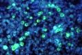 | Dr. Paul M. Feorino (CDC) 1972 Leukemia cells containing the Epstein Barr virus Photomicrograph, FA staining technique Creative Commons - Wikipedia This media comes from the Centers for Disease Control and Prevention's Public Health Image Library (PHIL), with identification number #2984. |
| 18. |  | David Gregory & Debbie Marshall 2005 Blood clot forming over a wound Scanning electron micrograph, colour-enhanced Wellcome Collection David Gregory & Debbie Marshall, Wellcome Images (B0006127) A blood clot, with squamous tissue visible beneath. As a blood clot on a surface injury dries out it forms a protective scab over the wound allowing new skin to grow underneath. |
| 19. |  | Ivor Mason, KCL 2009 Compact bone Light microscopy Wellcome Collection Ivor Mason, KCL, Wellcome Images (B0007263) Compact bone, from human femur, transverse sections showing cross sections and arrangement of osteons and Haversian canals. Bone is made up of two types of tissue: the compact bone forms a shell around the spongy cancellous bone that makes up the marrow space in the centre. Compact bone provides strength and rigidity and is solid in appearance. It is composed of a layered matrix of organic substances and inorganic salts that form around an intricate network of vasculature called Haversian canals (named after British physician Clopton Havers), shown in this image in red. The Haversian canals surround blood vessels and nerve cells throughout the bone and together with the layers of compact bone, the canals form units called osteons. The tiny black spaces shown in this image are due to the loss of osteocyte cells (living bone cells) that have disappeared during processing leaving the holes within the bone. Air is often trapped inside these holes during specimen preparation, giving the cavities a dark appearance because of optical refraction. Wellcome Image Award winner 2009 |
| 20. |  | K. Hodivala-Dilke & M. Stone n.d. SEM of blood vessel in a melanoma - coloured Freeze fracture scanning electron micrograph, colour-enhanced Wellcome Collection K. Hodivala-Dilke & M. Stone, Wellcome Images (B0003655) A colour-enhanced, freeze-fracture scanning electron micrograph of a blood vessel that has grown into a melanoma and is providing nourishment to it. Numerous red blood cells and three white blood cells can be seen within the blood vessel. |
| 21. |  | Freya Mowat 2005 Blood vessels emerging from the optic disc Confocal micrograph Wellcome Collection Freya Mowat, Wellcome Images (B0006228) Blood vessels in the retina emerging from the optic disc (black). The optic disc is the area of the retina where the optic nerve and retinal blood vessels leave the back of the eye. No light receptor cells are present in this area making it a blind spot. The endothelial cells are stained red and the vascular contents green. Surrounding cell nuclei are stained blue. |
| 22. |  | Matthieu Deuté 2006, 19 September Nematods from filets of codfish, deboned and dewormed in a fish market. Québec. Photomicrograph (?) Creative Commons - Wikipedia |
| 23. |  | Unknown creator 1974 Tapeworm [Taenia solium proglotid] Photomicrograph Creative Commons - Wikipedia Under a very low magnification of only 8X; this photomicrograph revealed some of the ultrastructural morphology exhibited by three Taenia solium proglottids. Proglottids of Taenia spp. Gravid proglottids are longer than wide and the two species, T. solium and T. saginata, differ in the number of primary lateral uterine branches: T. solium contains 7-13 lateral branches and T. saginata 12-30 lateral branches. Adults of Taenia spp. can reach a length of 2-8 meters, but the hooked scolex, located at the head region, is only 1-2 millimeters in diameter. Taeniasis is the infection of humans with the adult tapeworm of Taenia saginata or Taenia solium. Humans are the only definitive hosts for these two tapeworm species. This media comes from the Centers for Disease Control and Prevention's Public Health Image Library (PHIL), with identification number #1418. |
| 24. |  | Phillip D. Cartwright 1984, 1 December A dog flea under polarised light Photomicrograph Creative Commons - Wikipedia |
| 25. |  | MOutty (Wikipedia user id) 2010, 5 January Dolphin Foetus [Musée d'histoire naturelle de Bruxelles] Digital image Creative Commons - Wikipedia |
| 26. |  | Dr. Hannes Grobe 2010 Mummified cat (Senckenberg Naturmuseum, Frankfurt, Germany) Digital image Creative Commons - Wikipedia |
| 27. |  | Dr. Hannes Grobe 2005, 7 April Fluorescent minerals Under UV-A, UV-B and UV-C light Creative Commons - Wikipedia |
| 28. |  | Dr. Hannes Grobe 2005, 7 April Fluorescent minerals [key] Under UV-A, UV-B and UV-C light Creative Commons - Wikipedia Description of fluorescent minerals from page Fluorescence:
|
| 29. |  | Unknown creator 1995, February The Hubble Space Telescope imaged this view in February 1995. The arcing, graceful structure is actually a bow shock about half a light-year across, created from the wind from the star L.L. Orionis colliding with the Orion Nebula flow. Hubble telescope Creative Commons - Wikipedia |
| 30. |  | Unidentified photographer / artist n.d. Coronal loops Digital image NASA NASA, NASA Solar System Collection, UID: SPD-SLRSY-191, Image ID: 227136 Extending above the photosphere or visible surface of the Sun , the faint, tenuous solar corona can't be easily seen from Earth, but it is measured to be hundreds of times hotter than the photosphere itself. What makes the solar corona so hot? Astronomers have long sought the source of the corona's heat in magnetic fields which loft monstrous loops of solar plasma above the photosphere. Still, new and dramatically detailed observations of coronal loops from the orbiting TRACE satellite are now pointing more closely to the unidentified energy source. Recorded in extreme ultraviolet light, this and other TRACE images indicate that most of the heating occurs low in the corona, near the bases of the loops as they emerge from and return to the solar surface. The new results confound the conventional theory which relies on heating the loops uniformly. This tantalizing TRACE image shows clusters of the majestic, hot coronal loops which span 30 or more times the diameter of planet Earth. |
| 31. | 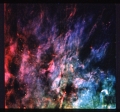 | Unidentified photographer / artist 1990, 4 October Window-Curtain - Structure of the Orion Nebula Revealed by NASA's Hubble Space Telescope Digital image NASA NASA Hubble Space Telescope Collection, UID: SPD-HUBBLE-STScI-199 0-26a Recent images made with the Wide Field Camera on NASA's Hubble Space Telescope have revealed the structure of a thin sheet of gas located at the edge of the famous "Great Nebula" in Orion, an estimated 1500 light years from Earth. Astronomers, who compare the appearance of this sheet of gas with that of a rippled window curtain, report that this emission traces the boundary between the hot, diffuse interior of the nebula and an adjacent dense cool cloud. The sheet is seen in light emitted by atoms of gaseous sulfur (shown in red in the photograph). This emission is strongest under conditions which are intermediate between those in the interior of nebula and those in the dense cloud. The sulfur emission is seen to break into filamentary and clumpy structures with sizes down to the limit of what the telescope can show. In contrast, emission from gaseous oxygen and hydrogen (shown as blue and green, respectively) is favored in the interior of the nebula itself, and is distributed much more smoothly in the image. The Orion Nebula is a "stellar nursery" - a region where new stars are forming out of interstellar gas. The emission from the nebula is powered by the intense ultraviolet light from a cluster of particularly hot and luminous stars. The sulfur emission seen in the photograph is coming from the region where the light from these stars is "boiling off" material from the face of the dense cloud. This is the very cloud from which the hot stars formed, and is known to harbor additional ongoing star formation. Astronomers say that this is a good example of a case where, despite the spherical aberration that has hobbled many of the scientific programs which the Hubble Space Telescope was expected to carry out, the telescope can still be used to obtain scientifically interesting data with clarity far exceeding that normally possible from the ground. The Wide Field/Planetary Camera was designed and built by the Jet Propulsion Laboratory, which is operated by the California Institute of Technology. |
| 32. |  | Cassini Orbiter 2007, 17 July Rhea - Icy Profile Imaging Science Subsystem - Narrow Angle NASA NASA/JPL/Space Science Institute, PIA10494 The Cassini spacecraft looks toward Rhea's cratered, icy landscape with the dark line of Saturn's ringplane and the planet's murky atmosphere as a background. Rhea is Saturn's second-largest moon, at 1,528 kilometers (949 miles) across. This view looks toward the unilluminated side of the rings from less than a degree above the ringplane. Images taken using red, green and blue spectral filters were combined to create this natural color view. The images were acquired with the Cassini spacecraft narrow-angle camera on July 17, 2007 at a distance of approximately 1.2 million kilometers (770,000 miles) from Rhea. Image scale is 7 kilometers (5 miles) per pixel. |
| 33. |  | Unidentified photographer / artist n.d. Halo of the Cat's Eye Digital image Source requested R. Corradi (Isaac Newton Group), D. Goncalves (Inst. Astrofisica de Canarias) The Cat's Eye Nebula (NGC 6543) is one of the best known planetary nebulae in the sky. Its haunting symmetries are seen in the very central region of this stunning false-color picture, processed to reveal the enormous but extremely faint halo of gaseous material, over three light-years across, which surrounds the brighter, familiar planetary nebula. Made with data from the Nordic Optical Telescope in the Canary Islands, the composite picture shows emission from nitrogen atoms as red and oxygen atoms as green and blue shades. Planetary nebulae have long been appreciated as a final phase in the life of a sun-like star. Only much more recently however, have some planetaries been found to have halos like this one, likely formed of material shrugged off during earlier active episodes in the star's evolution. While the planetary nebula phase is thought to last for around 10,000 years, astronomers estimate the age of the outer filamentary portions of this halo to be 50,000 to 90,000 years. This image was the "NASA - Astronomy Picture of the Day" - 2002, September 4. |
| 34. |  | Unidentified photographer / artist 2000, 8 August Mt. St. Helens ASTER NASA NASA/GSFC/METI/ERSDAC/JAROS, and U.S./Japan ASTER Science Team This ASTER image of Mt. St. Helens volcano in Washington was acquired on August 8, 2000 and covers an area of 37 by 51 km. Mount Saint Helens, a volcano in the Cascade Range of southwestern Washington that had been dormant since 1857, began to show signs of renewed activity in early 1980. On 18 May 1980, it erupted with such violence that the top of the mountain was blown off, spewing a cloud of ash and gases that rose to an altitude of 19 kilometers. The blast killed about 60 people and destroyed all life in an area of some 180 square kilometers (some 70 square miles), while a much larger area was covered with ash and debris. It continues to spit forth ash and steam intermittently. As a result of the eruption, the mountain's elevation decreased from 2,950 meters to 2,549 meters. The image is centered at 46.2 degrees north latitude, 122.2 degrees west longitude. |