Luminous-Lint - for collectors and connoisseurs of photography Register
Subscribe
Login
Photographers:
Connections:
Getting around...
| Home > Contents > Virtual exhibits
Explore subjects throughout photohistory.
Register and see for yourself...
Scientific: Contemporary
Title Carousel Lightbox Checklist
   
Unknown creator, 2007 (uploaded), DNA molecule, Rendered image, software generated, Creative Commons - Wikipedia, LL/42339
Unknown creator, 2006, 21 November, DNA repair, Rendered image, software generated, Creative Commons - Wikipedia, LL/42340
Kieran Maher, 2006, 30 October, 3D VR of a CT scan: orthogonal cropping frame, OsiriX, Creative Commons - Wikipedia, LL/42341
Milorad Dimic MD (Intermedichbo, Nis, Serbia), 2009, 16 August, Morbus Buerger, CT angiogram, Creative Commons - Wikipedia, LL/42342
Unknown creator, 2009, 27 October, This is a computer-enhanced fMRI scan of a person who has been asked to look at faces. The image shows increased blood flow in the part of the visual cortex that recognizes faces., Computer-enhanced fMRI scan, Creative Commons - Wikipedia, LL/42343
Fuchs, Daniel & Geo, n.d., Fetus, 8 months, with tumor, [Conserving Humans], Cibachrome print, Stephen Bulger Gallery, LL/2120
Ed Uthman, 2000 (ca), Human Embryo (7th week of pregnancy), Colour film, Creative Commons - Wikipedia, LL/42354
Unidentified photographer / artist, n.d., A scoliotic spine, National Museum of Health and Medicine, LL/6972
Unidentified photographer / artist, n.d., Egg surrounded by sperm, National Museum of Health and Medicine, LL/6973
Unidentified photographer / artist, n.d., The new M2A Capsule, a pill endoscope, created by Given Imaging Ltd, National Museum of Health and Medicine, LL/6971
Unidentified creator(s), n.d., Section through Visible Human Male - head, including cerebellum, cerebral cortex, brainstem, nasal passages (from Head subset), Color Cryosection, U.S. National Library of Medicine, Visible Human Project, LL/6887
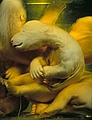
Unidentified creator(s), n.d., Section through Visible Human Male - abdomen, including large and small intestines, spinal column, musculature, subcutaneous fat (from Abdomen subset), Color Cryosection, U.S. National Library of Medicine, Visible Human Project, LL/6889
Fuchs, Daniel & Geo, n.d., Polar Bears, [Conserving Animals], Cibachrome print, Stephen Bulger Gallery, LL/2123
Unidentified photographer, 2010 (or earlier), Rat RPE layer, Colour image, National Institutes Of Health, LL/42571
Unidentified photographer, 2010 (or earlier), Heart, Colour image, National Institutes Of Health, LL/42572
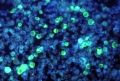
Dr. Paul M. Feorino (CDC), 1972, Leukemia cells containing the Epstein Barr virus, Photomicrograph, FA staining technique, Creative Commons - Wikipedia, LL/42348
David Gregory & Debbie Marshall, 2005, Blood clot forming over a wound, Scanning electron micrograph, colour-enhanced, Wellcome Collection, LL/36804
Ivor Mason, KCL, 2009, Compact bone, Light microscopy, Wellcome Collection, LL/36805
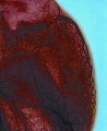
K. Hodivala-Dilke & M. Stone, n.d., SEM of blood vessel in a melanoma - coloured, Freeze fracture scanning electron micrograph, colour-enhanced, Wellcome Collection, LL/36806
Freya Mowat, 2005, Blood vessels emerging from the optic disc, Confocal micrograph, Wellcome Collection, LL/36807
Matthieu Deuté, 2006, 19 September, Nematods from filets of codfish, deboned and dewormed in a fish market. Québec., Photomicrograph (?), Creative Commons - Wikipedia, LL/42345
Unknown creator, 1974, Tapeworm [Taenia solium proglotid], Photomicrograph, Creative Commons - Wikipedia, LL/42346
Phillip D. Cartwright, 1984, 1 December, A dog flea under polarised light, Photomicrograph, Creative Commons - Wikipedia, LL/42347
MOutty (Wikipedia user id), 2010, 5 January, Dolphin Foetus [Musée d'histoire naturelle de Bruxelles], Digital image, Creative Commons - Wikipedia, LL/42352
Dr. Hannes Grobe, 2010, Mummified cat (Senckenberg Naturmuseum, Frankfurt, Germany), Digital image, Creative Commons - Wikipedia, LL/42353
Dr. Hannes Grobe, 2005, 7 April, Fluorescent minerals, Under UV-A, UV-B and UV-C light, Creative Commons - Wikipedia, LL/42349
Dr. Hannes Grobe, 2005, 7 April, Fluorescent minerals [key], Under UV-A, UV-B and UV-C light, Creative Commons - Wikipedia, LL/42350
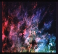
Unknown creator, 1995, February, The Hubble Space Telescope imaged this view in February 1995. The arcing, graceful structure is actually a bow shock about half a light-year across, created from the wind from the star L.L. Orionis colliding with the Orion Nebula flow., Hubble telescope, Creative Commons - Wikipedia, LL/42344
Unidentified photographer / artist, n.d., Coronal loops, Digital image, NASA, LL/36971
Unidentified photographer / artist, 1990, 4 October, Window-Curtain - Structure of the Orion Nebula Revealed by NASA's Hubble Space Telescope, Digital image, NASA, LL/36976
Cassini Orbiter, 2007, 17 July, Rhea - Icy Profile, Imaging Science Subsystem - Narrow Angle, NASA, LL/37367
Unidentified photographer / artist, n.d., Halo of the Cat's Eye, Digital image, Source requested, LL/37000
Unidentified photographer / artist, 2000, 8 August, Mt. St. Helens, ASTER, NASA, LL/37365
   
Title Carousel Lightbox Checklist

Terms and conditions • Copyright • Privacy • Contact me
Contributors retain copyright over their submissions
In using this website you agree to the Terms and Conditions
© Alan Griffiths - Luminous-Lint 2025