Contents
| This theme includes example sections and will be revised and added to as we proceed. Suggestions for additions, improvements and the correction of factual errors are always appreciated. Status: Collect > Document > Analyse > Improve | Introduction to photomicroscopy 381.01 Scientific > Introduction to photomicroscopy
Early photographers needed to have an undertanding of the sciences of chemistry and optics. The early camera makers were frequently opticians and the makers of scientific instruments and given this it is no surprize that they understood telescopes and microscopes. Within a few years of the announcement of photography in 1839 it was being used to record microscopic objects for scientific study.
- William Henry Fox Talbot (1800-1877), one of the inventors of photography and a remarkable polymath, took photomicrographs of plant and insect specimens using a solar microscope.
- John Benjamin Dancer (1812-1887) in 1840 showed a daguerreotype of a fly at the Liverpool Mechanics' Institution and the following year showed daguerreotypes taken with a microscope. By 1851 he was taking wet collodion photomicrographs and the following year selling microscopic portraits of Queen Victoria and the Lord's Prayer.
- John William Draper (1811-1882) took a photomicrograph (ca. 1850) on a daguerreotype of a Fly's proboscis (Smithsonian, National Museum of American History, Division of Information Technology and Society, Photographic History Collection)
- Wilson A. Bentley (1865-1931) could be described as a person with a fixation for the structure of snowflakes - in 1885 he attached a camera to a microscope and was able to create high quality albumen prints showing their different crystalline structure. In 1903 he supplied prints of his research to the Smithsonian in Washington. Interestingly James Glaisher, a President of the Royal Photographic Society, had preceded Wilson A. Bentley and had taken photomicrographs of snowflakes in 1855.
Photomicroscopists 381.02 Scientific > Henry Fox Talbot: Photomicrographs About this photographer | Photographs by this photographer
381.03 Scientific > John William Draper: Photomicrographs About this photographer | Photographs by this photographer
381.04 Scientific > Adolphe Bertsch: Photomicrographs About this photographer | Photographs by this photographer
381.05 Scientific > Joseph Janvier Woodward: Photomicrographs About this photographer | Photographs by this photographer
In 1862, during the still-ongoing American Civil War, Joseph Janvier Woodward was selected for the US War Department‘s newly-founded medical research facility, the Army Medical Museum. Woodward‘s duties included management of the Museum‘s medical work and microscopy as well as production of the three medical volumes for the massive Medical and Surgical History of the War of the Rebellion (1870-1888) whose final volume was published four years after his death. (Burns, 1983, p.1463)
In 1869 Woodward became the first microscopist to successfully resolve the 19th band of Norbert‘s 19-band test plate, created as test object for the resolution of period microscopes. While it was not understood at the time, Woodward was thereby approaching the absolute theoretical limits of the optical microscope, and the subsequent 20-band test-plates produced by Norbert contained rulings only capable of resolution by methods such as the electron microscope. (Turner, 1980, p.170)
Woodward‘s work at the Army Medical Museum involved extensive documentation through microphotography where again his work was on the frontier of period science. At the Museum he implemented important advances in microscopes, light-sources, tissue-dying techniques and integration of photography with microscopy, turning America into a world-leader in microphotography (Burns, 1983, p.1463). Not inappropriately Woodward has been called "the most skillful microphotographer of his age" (Frison 1954, p.120).
Woodward published several pamplets illustrating his advances with the microscope, often accompanied by superb 6 x 6 inch (16x16 cm) albumen prints of his photomicrographs on special gilt-bordered mounts. These typically measure 14 x 11 inches (35x28 cm) and bear the imprint "War Department, Surgeon General‘s Office, Army Medical Museum" ... "By J. J. Woodward".
Nine such plates were intended to accompany his Report to the Surgeon General of the United States Army on an Improved Method of Photographing Histological Preparations by Sunlight (1871), including Striated Muscular Fibres of Mouse and Small Artery and Capillaries from Lung of Frog. The plates bear pasted-on print labels identifying each subject as well as its magnification, series number, and other notations.
Woodward‘s Army Medical Museum plates were also issued in reference volumes and were presumably available to researchers individually. Thus, Woodward‘s photomicrograph of a Head Louse is found on his standard gilt War Department mount but with manuscript notations of subject and magnification rather than a printed label, perhaps indicating that it was issued one-off and not as part of a series.
Woodward's beautiful microscopic images present a jarring intersection between the onset of advanced scientific technology and an era we more often associate with muskets and hardtack.
[Courtesy of Christopher Wahren] 381.06 Scientific > Frederick H. Evans: Photomicrographs About this photographer | Photographs by this photographer
381.07 Scientific > Wilson A. Bentley: Photomicrographs of snowflakes About this photographer | Photographs by this photographer
381.08 Scientific > Richard Neuhauss: Photomicrographs of snowflakes About this photographer | Photographs by this photographer
381.09 Scientific > Laure Albin-Guillot (1879-1962): Micrographie Décorative About this photographer | Photographs by this photographer
The patterns shown were so fantastic that parallels were seen between them and contemporary abstract art and some of the works of Laure Albin-Guillot (1879-1962) and the Sorbonne geology professor H. Ragot were published in The Illustrated London News (May 1931). 381.10 Scientific > Alfred Ehrhardt: Photomicroscopy About this photographer | Photographs by this photographer
381.11 Scientific > Carl Strüwe: Photomicrographs About this photographer | Photographs by this photographer
Carl Heinrich Jakob Strüwe (1898–1988) was a professional graphic designer who spent his career as an employee of a graphics company in Bielefeld, Germany. As an artist and photographer he was self taught but he exhibited and published before and after the Second World War in first class galleries and museums. In the 1950s he became a member of the movement of Subjective Photography of Otto Steinert in West Germany and participated in his exhibitions and publications. He was an innovative photographer and a pioneer in the art of microphotography. Subjects for photomicroscopy
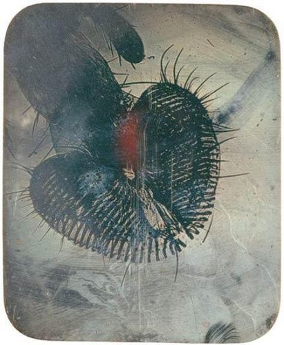
 381.12 Scientific > Photomicrographs of insects
381.13 Scientific > Rose-Lynn Fisher: Bee About this photographer | Photographs by this photographer
Reflections
Rose-Lynn Fisher
Photographs of a honeybee through a scanning electron microscope reveal a realm of design and function that stretches our sense of scale and wonder to another order of magnitude and brings science to the threshold of art. With highly magnified views of the bee’s eyes, antennae, wings, legs, hair, and abdomen through the perspective of microscopy, these images present a new frontier right here in our everyday world. In the process of exploring a bee under a microscope, we enter a realm of design, structure and pattern at an astonishing level of detail; and as the magnifications increase, the integrity of form is continuously revealed. When our sense of scale has no frame of reference, the micro and macro worlds seem interchangeable. Our familiar context confounded, the interplay of observation and imagination can inspire new ideas, connections, and applications. Seeing what exists at the micro level naturally extends to a more sensitized awareness of what is all around us in the visible and invisible worlds. Considering the endless structures and forms that comprise a little bee at higher and higher magnifications, we get a hint of the amazing, unending complexity of nature all around us. This can be startling and inspiring, and my hope is to foster deeper curiosity, greater appreciation, awe and marvel for the honeybee. After looking up close at the honeybee, one can never think of this tiny amazing creature in the same way again.
Rose-Lynn Fisher Bee (Princeton Architectural Press, 2010).
Of the ten million or so different species of insects on our planet, none is more fascinating than the honeybee. one of the oldest forms of animal life still in existence from the Neolithic age, bees have been worshipped and mythologized since the beginning of human history. Known popularly for their industriousness (“as busy as a bee”) and highly valued for their role in agricultural pollination (every third bite we take depends on them), bees are now kept by a quarter-million beekeepers in the united states alone, and millions more around the world.
Honeybees were the first creatures examined by seventeenth-century scientists whose primitive microscopes suggested a complex system of construction. Now, magnified hundreds to thousands of times with a scanning electron microscope, honeybees appear as architectural masterpieces—an elegant fusion of form and function.
Melding art and science, photographer Rose-Lynn Fisher puts this modern tool to creative use in order to reveal the microscopic majesty of these natural wonders. BEE presents sixty astonishing photographs of honeybee anatomy in magnifications ranging from 10x to 5000x. rendered in stunning detail, Fisher’s photographs uncover the strange beauty of the honeybee’s pattern, form, and structure. Comprising 6,900 hexagonal lenses, their eyes resemble the structure of a honeycomb. the honeybee’s proboscis—a strawlike appendage used to suck nectar out of flowers, folds resembles a long, slender hairy tongue. Its six-legged exoskeleton is fuzzy with hairs that build up a static charge as the bee flies in order to electrically attract pollen. wings clasp together with tiny hooks and a double-edged stinger resembles a serrated hypodermic needle. the honeybee’s three pairs of segmented legs are a revelation, with their antennae cleaners, sharp-pointed claws, and baskets to carry pollen to the hive. these visual discoveries, made otherworldly through Fisher’s lens, expand the boundaries of our thinking about the natural world and stimulate our imaginations. BEE features a foreword by nature writer and New York Times editorial board member Verlyn Klinkenborg. 381.14 Scientific > Photomicrographs of botany
Copying paintings and documents 381.15 Scientific > Henry Hering - Alfred Reeves: Photograph. The Kings and Queens of England
381.16 Scientific > W. & F. Langenheim: The Lord's Prayer About this photographer | Photographs by this photographer
alan@luminous-lint.com |
General reading 1870-88, Medical and Surgical History of the War of the Rebellion (1865-65), (Washington, Gov’t print. off.) [Three volumes. Includes work by Joseph Janvier Woodward] [Δ] American Stereoscopic Company (Philadelphia), 1861, Catalogue of Langenheim's New and Superior Style of Colored Photographic Magic Lantern Pictures - Also a Catalogue of Langenheim's Stereoscopic Pictures on Glass and Paper, and Microscopic Photographs of a Superior Quality, (E. Ketterlinus, printer) [Δ] Girard, Jules, 1869, La Chambre Noire et le Microscope, (Paris: F. Savy) [Δ] Keller, Corey, 2008, Brought to Light: Photography and the Invisible, 1840-1900, (Yale University Press) isbn-10: 0300142102 isbn-13: 978-0300142105 [Δ] Libbrecht, Kenneth, 2007, The Art of the Snowflake: A Photographic Album, (Voyageur Press) isbn-10: 0760329974 isbn-13: 978-0760329979 [Δ] Scott, Jean, 2002, Stanhopes: A Closer View - A History and Handbook for Collectors of Microphotographic Novelties, (Greenlight Publishing) isbn-10: 1897738099 isbn-13: 978-1897738092 [Δ] Seiler, Carl, 1879, ‘Photography as an Aid to Microscopical Investigations‘, Proceedings of the American Society of Microscopists, vol.1, pp.59-62 [Δ] Readings on, or by, individual photographers René Patrice Proudhon Dagron Dagron, R.P.P., 1864, ‘Microscopic photography‘, British Journal of Photography, pp.402 [Δ] Dagron, R.P.P., 1864, Traité de Photographie Microscopique, (Paris) [Δ] John Benjamin Dancer Arden, L.L., 1960, John Benjamin Dancer: The Originator of Microphotography, (London: The Library Association) [Δ] Bracegirdle, Brian & McCormick, James B., 1993, The Microscopic Photographs of J.B. Dancer, (Chicago: Science Heritage Ltd) isbn-10: 0940095106 isbn-13: 978-0940095106 [Δ] Alfred Ehrhardt Derenthal, Ludger & Stahl, Christiane (eds.), 2010, Mikrofotografie - Schönheit jenseits des Sichtbaren (Microphotography - Beauty beyond the Visible World), (Hatje Cantz Verlag) isbn-13: 978-3775726788 [Δ] Ruge, Werner, 1939, Die Melodie des Lebens Ein Bildbuch aus der Zeit der Wende abendländischen Denkens, (Leipzig: Reclam Verlag) [Mit 40 mikroskopischen Aufnahmen von Alfred Ehrhardt] [Δ] Lennart Nilsson Forsell, Jacob (ed.), 2002, Lennart Nilsson: Images of His Life, (Stockholm: Bokförlaget Max Ström) [Δ] Carl Strüwe Jäger, Gottfried, 1982, Carl Strüwe, Das fotografische Werk, (Edition Marzona) [Δ] Strüwe, Carl, 1955, Carl Strüwe: Formen des Mikrokosmos. Gestalt und Gestaltung einer Bilderwelt, (Munich: Prestel-Verlag) [Δ] Strüwe, Carl, 1986, Carl Strüwe: Hohenstaufen in Italien. Bilder und Worte, (Bielefeld: Edition Jesse) [Δ] Joseph Janvier Woodward Woodward, J.J., 1873, November, U.S. Army Smithsonian Miscellaneous Collections, 262, The Toner Lectures, Washington, U.S.A., Lecture I: "On the Structure of Cancerous Tumours, and the Mode in which Adjacent Parts are Invaded." [Δ] Woodward, Joseph Janvier, 1871, Report to the Surgeon General of the United States Army on an Improved Method of Photographing Histological Preparations by Sunlight, (Washington D.C.) [Δ] If you feel this list is missing a significant book or article please let me know - Alan - alan@luminous-lint.com Laure Albin-Guillot (1879-1962) • Alois Auer (1813-1869) • Wilson A. Bentley (1865-1931) • Adolphe Bertsch • René Patrice Proudhon Dagron (1818-1900) • John Benjamin Dancer (1812-1887) • Alfred Donné (1801-1878) • John William Draper (1811-1882) • Alfred Ehrhardt (1901-1984) • Frederick H. Evans (1853-1943) • Claudia Fährenkemper • Albert Fernique (1841-1898) • William Towler Kingley (1815-1916) • Robert Koch (1843-1910) • Frederick Langenheim (1809-1879) • W. & F. Langenheim • William Langenheim (1807-1874) • Richard Neuhauss • Adolphe Neyt (1830-1893) • Jan Evangelista Purkinje (1787-1869) • Alfred Reeves (1829-1907) • George Shadbolt (1830-1901) • John Charles Stovin (1814-1896) • Carl Strüwe (1898-1988) • Henry Fox Talbot (1800-1877) • Joseph Janvier Woodward (1833-1884) | Home > Themes > Scientific > Photomicroscopy
|